
Nexus of Pathophysiology and Therapeutics (NPT) is an international, peer-reviewed journal publishing research in pathophysiology, biomedical sciences, pharmacology, and therapeutics, with a focus on disease mechanisms and clinical applications.
Document Type : Systematic Review
Authors
1 Department of Pharmacotherapy, School of Pharmacy. Mazandaran University of Medical Sciences, Iran.
2 Medical Plants Research Center, Basic Health Sciences Institute, Shahrekord University of Medical Sciences, Shahrekord, Iran.
3 Department of Pharmacology, School of Medicine, Shahrekord University of Medical Sciences, Shahrekord, Iran.
4 Department of Pharmacology, School of Medicine, Tehran University of Medical Sciences, Tehran, Iran.
5 Experimental Medicine Research Center, Tehran University of Medical Sciences, Tehran, Iran.
Graphical Abstract
Keywords
1. Introduction
Acetaminophen (paracetamol) is one of the most widely used analgesic and antipyretic drugs worldwide (1). At recommended doses, it has a well-established safety profile and remains an important component of pain and fever management (2). However, excessive exposure can cause severe liver injury and, in susceptible individuals, progress to acute liver failure (ALF) (3). Acetaminophen overdose is a major cause of acute liver failure in several Western countries and may result from either intentional overdose or unintentional exposure, including repeated use or combined administration of acetaminophen-containing products (4). Acetaminophen-induced liver injury involves multiple interacting pathways, including oxidative and endoplasmic reticulum (ER) stress, mitochondrial impairment, and cell death (5). The classical model established the importance of hepatic bioactivation, glutathione depletion, and covalent binding of the reactive metabolite N-acetyl-p-benzoquinone imine (NAPQI) to cellular proteins (6, 7). Subsequent research implicated mitochondrial dysfunction, oxidative stress, JNK signaling, apoptotic changes, and necrotic cell death as important components of acetaminophen-induced liver injury (APAP-induced liver injury; AILI) (8). More recent studies have highlighted adaptive responses, mitochondrial quality-control mechanisms, biomarkers of mitochondrial dysfunction, sterile inflammation, and hepatic regeneration (9).
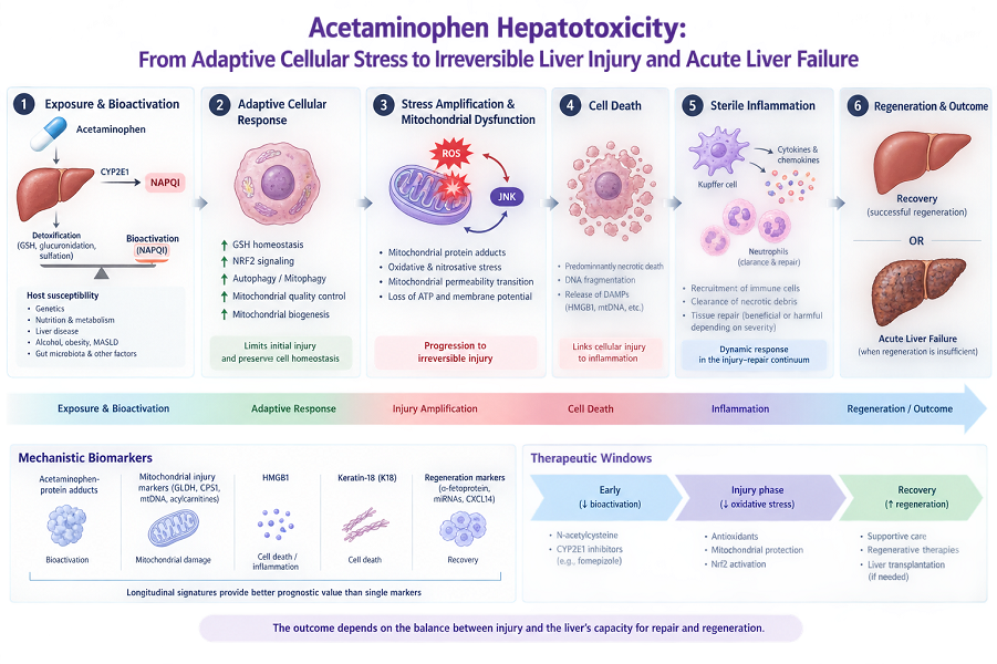
Acetaminophen hepatotoxicity is not a linear process in which NAPQI formation inevitably causes hepatocyte death (10). Its outcome depends on the interplay between the extent of liver injury and the liver’s capacity for effective repair and regeneration (11). When adaptive mechanisms are overwhelmed, mitochondrial dysfunction and cellular stress progress toward irreversible hepatocyte injury and death (12). Subsequent inflammation and regenerative responses can influence whether liver function recovers or progresses to acute liver failure (13). Mechanistic biomarkers of bioactivation, mitochondrial injury, and cell death may provide information beyond conventional aminotransferases and help define the evolving stage of injury. This Review integrates exposure, host susceptibility, adaptive responses, mitochondrial dysfunction, cell death, inflammation, regeneration, biomarkers, and stage-specific therapy into a dynamic injury–recovery framework to explain differences in biological and clinical outcomes after acetaminophen toxicity.
2. Exposure, Bioactivation, and the Initial Toxicological Challenge
The biological response to acetaminophen is strongly influenced by the characteristics of exposure, including dose, duration, and timing (14). Although acute overdose remains the most recognized pattern of paracetamol toxicity, the magnitude and pattern of exposure, including dose, duration, and repeated or staggered administration, can substantially influence the risk and severity of hepatotoxicity (15). Repeated supratherapeutic ingestion is particularly important because clinically significant hepatotoxicity can develop after repeated exposures that do not involve a large acute single overdose (15). Thus, exposure should be viewed as a dynamic input that determines the extent of metabolic challenge rather than as an isolated dose event. At therapeutic doses, most acetaminophen is eliminated through glucuronidation and sulfation. A smaller fraction undergoes oxidation by cytochrome P450 enzymes, principally CYP2E1, to generate the reactive electrophile N-acetyl-p-benzoquinone imine (NAPQI). Under normal conditions, NAPQI is efficiently conjugated with hepatic glutathione (GSH), promoting its detoxification and reducing its availability for covalent binding to cellular proteins (16). This balance between metabolic activation and detoxification enables the liver to handle therapeutic acetaminophen exposure without significant hepatocellular injury (17). Excessive exposure disrupts this balance by increasing NAPQI formation and depleting GSH required for its detoxification (18). Unneutralized NAPQI then forms covalent adducts with cellular proteins (19). Although protein adducts can provide evidence of acetaminophen bioactivation, their biological significance extends beyond exposure assessment because NAPQI-mediated protein modification can cause protein dysfunction, particularly in mitochondria, thereby disrupting the electron transport chain, increasing oxidative stress, and ultimately contributing to hepatocellular injury (19). The extent of protein adduct formation therefore reflects the balance between reactive-metabolite generation and the liver’s capacity to detoxify or neutralize these reactive species (20). Excessive NAPQI formation in the setting of GSH depletion should therefore be regarded as an initiating toxicological challenge rather than as synonymous with irreversible hepatotoxicity (21). The critical consequence of excessive bioactivation is the formation of protein adducts, with mitochondrial protein adduction serving as an important initiating event that triggers mitochondrial oxidant stress and subsequent amplification of oxidative and nitrosative stress (22). Whether this initial challenge is successfully contained or progresses toward substantial cellular injury depends on the hepatic capacity to respond to the resulting stress (23). The extent of NAPQI formation is only one determinant of toxicity. The ultimate outcome also depends on whether hepatocytes can withstand the resulting cellular and mitochondrial stress (23).
3. Host Susceptibility and Hepatic Resilience
The hepatic response to acetaminophen is influenced not only by the dose and exposure regimen but also by host susceptibility and biological factors (24). The severity of drug-induced liver injury can vary among individuals because susceptibility is influenced by metabolic capacity, nutritional status, underlying liver disease, concomitant exposures, and other host-related factors (25). Host susceptibility may therefore be viewed, at least in part, as a modifier of hepatic resilience, the capacity of the liver to withstand metabolic stress while preserving redox and bioenergetic homeostasis (26). Underlying metabolic dysfunction may influence hepatocellular resilience by altering cellular stress responses and hepatic detoxification capacity (27). Malnutrition and starvation have been proposed to reduce hepatic glutathione availability and alter acetaminophen metabolism, potentially increasing susceptibility to toxicity, particularly during repeated exposure; however, the clinical contribution of these factors remains uncertain (28). Chronic alcohol exposure can alter CYP2E1 activity and hepatic redox status, although its effect cannot be attributed to bioactivation alone (29). Similarly, obesity and pre-existing metabolic dysfunction-associated steatotic liver disease (MASLD) may increase susceptibility to acetaminophen-induced hepatic injury, potentially through disturbances in hepatic lipid handling and mitochondrial fatty acid oxidation (30). Importantly, experimental findings in obesity and fatty liver disease are not entirely consistent (31). Increased CYP2E1 activity may favor APAP bioactivation to NAPQI, while concurrent variation in alternative metabolic and detoxification pathways may modify the overall balance between bioactivation and clearance (32). These conditions should therefore be regarded as complex biological modifiers rather than uniform predictors of toxicity (33). Genetic variation represents an additional source of interindividual variability in acetaminophen disposition, with UGT1A6 variants influencing drug exposure and elimination (34). Differences in enzymes involved in acetaminophen metabolism can alter the relative contribution of bioactivation and detoxification, while variation in endogenous defense systems may influence the response to the resulting cellular stress (35). Among these systems, nuclear factor erythroid 2-related factor 2 (NRF2) is particularly relevant because it regulates the expression of genes involved in antioxidant defense and detoxification of reactive electrophiles, thereby contributing to cellular protection (36). Experimental evidence suggests that NRF2 activity contributes to the cellular defense against APAP-induced stress and may influence susceptibility to subsequent hepatotoxic injury (37).
The host environment may also shape susceptibility to APAP-induced liver injury, as suggested by the effects of excessive alcohol intake and fasting (38). Gut microbial metabolites have emerged as potential modulators of acetaminophen susceptibility, with different microbial products capable of altering hepatic metabolic and stress responses (39). This emerging gut–liver connection suggests that susceptibility is not determined exclusively by intrinsic hepatic processes but can also be shaped by systemic metabolic, microbial, immune, and other extrahepatic factors (40). Overall, host susceptibility provides the biological context that determines how acetaminophen exposure is handled and translated into hepatic injury (40). Individual susceptibility to clinically relevant APAP-induced liver injury is influenced by multiple interacting factors including genetic variation and the biological context in which exposure occurs. No single factor should be interpreted as an isolated determinant of outcome (41). Their significance therefore lies not in their individual effects, but in how their interactions collectively influence hepatic susceptibility, injury severity, and the capacity for recovery (42). Once the initial toxicological challenge has been established, the subsequent course of APAP-induced liver injury is shaped not only by the extent of cellular damage but also by the ability of hepatocytes to mount adaptive responses that modulate inflammation and support recovery (43).
4. From Initial Cellular Stress to Adaptive Response
Acetaminophen-protein adduct formation is not synonymous with hepatocyte death, as mitochondrial dysfunction and ROS accumulation are required for progression to cell death (44). Following the initial APAP-induced injury, surviving hepatocytes contribute to the regenerative response through proliferative and repair-associated mechanisms that support restoration of liver mass and function (11). The effectiveness of these responses is an important determinant of whether the initial stress remains contained or progresses toward more extensive injury (11). Autophagy is one of the principal mechanisms involved in this adaptive response. By delivering damaged proteins and organelles to lysosomes for degradation, autophagy can limit the accumulation of cellular components modified during acetaminophen exposure (45). Experimental studies indicate that impaired autophagy can exacerbate acetaminophen-induced liver injury, supporting a protective role for autophagy in limiting hepatic damage (46). Mitophagy provides a more selective form of quality control by removing dysfunctional mitochondria (47). Maintaining mitochondrial quality is particularly important because dysfunctional mitochondria can amplify oxidative stress and contribute to downstream molecular events leading to cell death, whereas their selective clearance through mitophagy can help restore cellular homeostasis (47).
NRF2 provides a transcriptional defense system that enhances cellular protection against oxidative stress by inducing protective and detoxification-related genes (48). Activation of Nrf2 promotes the expression of antioxidant and detoxification genes involved in cellular defense against oxidative and xenobiotic stress (49). Experimental evidence indicates that impaired NRF2 signaling can increase vulnerability to acetaminophen-induced cellular injury, whereas NRF2 activation enhances cytoprotective responses (50). Thus, NRF2 functions as an important regulatory component of the adaptive response rather than simply reflecting the presence of oxidative stress (51). Adaptive capacity also involves preserving mitochondrial function and limiting mitochondrial dysfunction (51). Surviving hepatocytes can modify mitochondrial morphology and activate mitochondrial biogenesis to replenish the functional mitochondrial pool after cellular stress (52). Together, autophagy and mitophagy contribute to cellular quality control by promoting the removal of damaged cellular components and dysfunctional mitochondria, thereby supporting mitochondrial function and limiting oxidative stress (53). Their importance may extend beyond immediate survival because preservation of viable hepatocytes can help maintain the cellular capacity required for subsequent tissue repair and regeneration (54). The early cellular response to acetaminophen toxicity attempts to contain the initial insult, while necrosis can trigger subsequent inflammatory and tissue-repair responses (54). Autophagy and mitophagy remove damaged components, NRF2 reinforces endogenous defense systems, and mitochondrial remodeling supports restoration of cellular function (55). These mechanisms do not eliminate the initial toxic challenge but can limit its consequences and preserve cellular homeostasis. However, when cellular stress becomes sustained or excessive, protective responses may become insufficient, while oxidative stress and mitochondrial dysfunction can progressively amplify hepatocellular injury in acetaminophen toxicity (56).
5. Stress Amplification and Mitochondrial Dysfunction
When the initial toxic challenge overwhelms cellular homeostatic capacity, acetaminophen-induced stress can become self-amplifying through interconnected ROS and Ca²⁺ signaling loops (57). Mitochondria occupy a central position in this process because they are not merely passive targets of injury but active amplifiers of cellular damage (57). Mitochondrial protein modifications, impaired respiratory function, reactive oxygen and nitrogen species, and stress-responsive signaling interact to progressively disrupt mitochondrial homeostasis (58). Mitochondrial protein adduct formation can impair mitochondrial respiration and promote electron leakage from the respiratory chain, contributing to mitochondrial superoxide generation (59). This oxidant signal contributes to activation of stress-responsive pathways, particularly c-Jun N-terminal kinase (JNK) (60). Following its activation by upstream stress-activated kinases, JNK translocates to mitochondria, where it further disrupts mitochondrial function and amplifies oxidative stress (60). This creates a positive feedback loop in which mitochondrial dysfunction increases oxidant stress, while JNK signaling further disrupts mitochondrial function (60).
Mitochondrial superoxide can react with nitric oxide to form peroxynitrite (ONOO−), a highly reactive species that can promote protein nitration and alter protein function (61). Experimental evidence supports an important role for mitochondrial oxidant and nitrosative stress in acetaminophen hepatotoxicity (62). The significance of this process is therefore not simply the generation of reactive species, but the establishment of an amplifying loop in which oxidant stress promotes JNK activation, while JNK-dependent mitochondrial dysfunction further intensifies mitochondrial oxidant stress (62). This helps explain why injury can progress even after the initial metabolic challenge has occurred. As mitochondrial dysfunction progresses, mitochondrial permeability transition (MPT) may emerge as an important downstream event contributing to further mitochondrial injury (63). Loss of mitochondrial membrane potential and impaired oxidative phosphorylation compromise ATP generation and cellular energy homeostasis (63). Mitochondrial swelling and structural alterations can impair mitochondrial function, limiting ATP production and compromising essential hepatocyte functions (64). At this stage, mitochondrial injury extends beyond a localized biochemical disturbance, compromising oxidative phosphorylation, ATP production, and overall cellular bioenergetics (64).
Importantly, early mitochondrial dysfunction may be amenable to cellular quality-control mechanisms, whereas sustained oxidative stress and JNK activation can promote further mitochondrial damage and functional failure (65). Thus, mitochondrial dysfunction may represent a critical transition point at which cellular stress progresses toward irreversible injury (65). The cellular outcome is closely linked to the preservation or restoration of mitochondrial function, including oxidative phosphorylation and the regulation of reactive oxygen species (66). Interactions of metallodrugs with cellular proteins and thiol-containing biomolecules can alter intracellular redox homeostasis and mitochondrial function, ultimately contributing to oxidative damage and cell death (67). Mitochondrial disruption can compromise oxidative phosphorylation and membrane integrity, promote oxidative stress, and ultimately drive metabolic collapse and cell death (67).
6. The Transition to Irreversible Injury and Cell Death
Early mitochondrial dysfunction does not necessarily result in irreversible hepatocyte death (9). During limited or transient mitochondrial stress, surviving cells may engage adaptive responses that preserve mitochondrial function and allow recovery from the initial insult (9). The critical transition occurs when mitochondrial injury becomes severe or persistent enough to exceed this compensatory capacity (23). At that point, loss of bioenergetic and structural homeostasis increasingly commits the hepatocyte to cell death (68). A late consequence of severe mitochondrial injury is the release of mitochondrial intermembrane-space proteins, particularly apoptosis-inducing factor (AIF) and endonuclease G (EndoG), followed by their translocation toward the nucleus (60). These events contribute to extensive nuclear DNA fragmentation. Although DNA damage is not an initiating event in acetaminophen hepatotoxicity, nuclear DNA fragmentation occurs downstream of severe mitochondrial dysfunction and permeability transition and is associated with progression toward regulated necrotic cell death (60). In this sense, extensive nuclear DNA fragmentation may mark a critical transition from cellular stress with adaptive potential to irreversible hepatocyte death (17).
The predominant mode of hepatocyte death after clinically relevant acetaminophen overdose is necrotic, with loss of plasma-membrane integrity and release of intracellular contents into the circulation (41). Because APAP-induced hepatocyte death is driven by a defined sequence of intracellular events, including mitochondrial damage and oxidative stress, rather than by nonspecific passive cellular destruction, it has been considered a form of regulated necrosis (41). This distinction is important when interpreting studies that identify molecular features associated with other forms of regulated cell death (69). Apoptosis, necroptosis, ferroptosis, and pyroptosis have all been investigated in acetaminophen hepatotoxicity (70). However, detection of an individual molecular marker should not be interpreted as definitive evidence of a specific or dominant cell-death pathway, particularly when multiple mechanisms may contribute to hepatocyte injury (71). The relative contribution of acetaminophen metabolic pathways can vary with dose, exposure conditions, tissue context, genetic background, and pharmacological modulation (72). Recent studies therefore emphasize that cell-death responses to acetaminophen are highly dependent on the experimental context, including the tissue examined and the molecular level of analysis (73). Ferroptosis illustrates this principle particularly well. Iron-dependent lipid peroxidation can contribute to hepatocyte injury under specific experimental conditions, but current evidence does not support ferroptosis as the dominant mechanism of cell death under pathophysiologically relevant conditions of acetaminophen overdose (74). Experimental manipulation of the cellular environment, particularly iron availability, can enhance iron-dependent lipid peroxidation and increase its contribution to cell injury (74). Such findings broaden the mechanistic landscape without replacing the established mitochondrial oxidant stress and necrotic pathway under typical conditions (75).
Taken together, excessive acetaminophen bioactivation initiates mitochondrial stress that, when sufficiently amplified, progresses to irreversible mitochondrial dysfunction, DNA fragmentation, and predominantly necrotic hepatocyte death (75). Alternative cell-death pathways may contribute under specific experimental conditions, but current evidence does not support considering them equivalent to the established central mechanisms of APAP toxicity (21). Necrotic hepatocyte death releases DAMPs, including HMGB1 and DNA fragments, which activate resident macrophages through innate immune receptors and initiate sterile inflammation (76). Thus, hepatocyte death links intracellular injury amplification to tissue-level inflammation, clearance, and subsequent regeneration (76).
7. Sterile Inflammation and the Injured Liver
Once hepatocyte death occurs, acetaminophen-induced injury can extend beyond the initially affected cells to neighboring hepatocytes (77). Cell death during APAP-induced liver injury releases damage-associated molecular patterns (DAMPs), including HMGB1, nuclear DNA fragments, mitochondrial DNA, and ATP (78). These signals can be sensed through receptors such as TLR4 and RAGE on hepatic immune cells, promoting inflammatory mediator production in the absence of an infectious pathogen (78). Sterile inflammation is therefore best viewed as a downstream response to APAP-induced hepatocyte injury rather than as the primary initiating mechanism of hepatotoxicity (79). DAMPs released from necrotic hepatocytes stimulate cytokine and chemokine production by resident Kupffer cells, thereby promoting the recruitment of circulating monocytes and neutrophils to the injured liver (80). Although neutrophils generate reactive oxygen species and other potentially injurious mediators, evidence for a major role in APAP-induced hepatocellular necrosis remains limited (81). Instead, recruited neutrophils may contribute to the clearance of necrotic debris and subsequent tissue repair (81). The presence of inflammatory cells in the injured liver should therefore not be interpreted automatically as evidence that inflammation is the primary cause of hepatocellular damage (82).
The functional consequences of sterile inflammation are strongly dependent on injury severity and disease stage (83). Following tissue injury, DAMP-mediated immune signaling promotes the recruitment of immune cells, facilitates clearance of necrotic cell debris, and modulates signaling between immune and parenchymal cells to support tissue repair (84). Macrophage populations can undergo functional changes during this process, providing signals that help organize the transition from tissue injury toward repair (84). In contrast, extensive or persistent hepatocyte loss can overwhelm these compensatory responses and become associated with impaired tissue recovery (85). Sterile inflammation may therefore be viewed as a dynamic component of the injury repair continuum, with its effects depending on the magnitude, timing, and cellular context of the response (85). Its significance depends on the extent of tissue injury, the timing of immune activation, and the functional phenotype of responding macrophages (86). This stage-dependent behavior is important when interpreting experimental findings, as inflammatory responses and leukocyte infiltration accompany active tissue injury, followed by cellular changes associated with tissue repair (86). The release of DAMPs therefore establishes an important biological link between hepatocyte death and the subsequent inflammatory and immune responses that shape liver injury (87). The regenerative outcome of liver injury depends on the severity of hepatocyte loss and the capacity of surviving hepatocytes to proliferate and restore liver function (88).
8. Regeneration, Recovery, and Acute Liver Failure
The extent of hepatocyte injury and death does not necessarily determine the final clinical outcome of acetaminophen hepatotoxicity (43). The liver has a remarkable capacity to regenerate hepatocytes after acute cellular injury, and effective regeneration may be an important component of recovery from severe hepatic injury and prevention of progression to liver failure (43). Following acetaminophen overdose, regenerative responses develop within a microenvironment shaped by necrosis, inflammatory signaling, and cellular stress (89). Consequently, successful recovery depends not only on the availability of mitogenic signals but also on the ability of surviving hepatocytes and non-parenchymal cells to respond to them (11). After moderate liver injury, regenerative activity increases in proportion to the extent of hepatic injury and can progressively restore liver mass and function as the injury regresses (11). Hepatocyte growth factor, epidermal growth factor, IL-6, and TNF-α contribute to signaling networks that promote hepatocyte proliferation and cell-cycle progression, whereas Wnt/β-catenin signaling regulates proliferative responses and liver regeneration following acetaminophen-induced injury (90). Macrophages, endothelial cells, hepatic stellate cells, and other non-parenchymal populations contribute to hepatic repair through diverse processes, including cellular proliferation, angiogenesis, and matrix remodeling (91). Regeneration is therefore a coordinated multicellular response rather than simply the proliferation of surviving hepatocytes (91). Importantly, reparative and regenerative programs can be engaged during the ongoing injury response, with endothelial, immune, and other non-parenchymal cells contributing to tissue restoration (91). Experimental studies have shown that primary hepatocytes can spontaneously organize into three-dimensional spheroid structures with features of functional liver architecture (92). Recent single-cell studies have revealed distinct hepatocyte populations and dynamic cellular states during liver regeneration, while also highlighting coordinated proliferation, matrix remodeling, and other multicellular processes involved in restoration of liver mass and function (93).
The critical transition toward acute liver failure occurs when regenerative capacity becomes insufficient relative to the magnitude or persistence of injury (94). Severe acetaminophen toxicity may leave regenerative pathways activated while simultaneously limiting the ability of surviving hepatocytes to respond (42). Persistent DNA damage, inflammatory signaling, cellular stress, and anti-proliferative pathways involving p53, p21, and transforming growth factor-β can promote cell-cycle arrest (42). In addition, hepatocyte injury and loss can alter the regenerative response and, when persistent, may contribute to pathological remodeling of the liver (95). Thus, regenerative failure may reflect disruption of the signaling pathways that normally support hepatocyte-driven liver regeneration (95). ALF cannot be explained solely by the extent of hepatocyte death; it also reflects loss of hepatic function and systemic complication (96). This may reflect impaired restoration of hepatic mass and function when ongoing tissue injury limits the regenerative response (97). When regeneration compensates for tissue loss, hepatic mass and function can recover, although the regenerated architecture may not fully reproduce the original anatomy (97). The liver has a remarkable capacity to regenerate after injury (97). However, factors related to the severity of injury and the underlying hepatic condition can impair this regenerative response and may contribute to hepatic dysfunction or failure (97). The outcome of acetaminophen toxicity can therefore be conceptualized as the interplay between hepatic injury and the capacity for repair and regeneration (98). Although the severity of the initial hepatic insult is an important determinant of outcome, subsequent repair and regeneration of the remaining liver may also substantially influence recovery (99). This perspective shifts attention from hepatocyte death alone toward the dynamic pathophysiological state of the injured liver and provides a biological basis for identifying biomarkers that reflect distinct phases of ongoing injury, inflammation, and regenerative recovery (100).
9. Therapeutic Windows and Stage-Specific Intervention
The temporal evolution of acetaminophen hepatotoxicity has important therapeutic implications, as molecular pathways that contribute to injury may differ across stages of disease progression (101). N-acetylcysteine (NAC) remains the established antidote for acetaminophen toxicity and is most effective when administered early, acting in part as a glutathione precursor to facilitate NAPQI detoxification (102). Its benefits may persist after liver injury has developed by limiting mitochondrial oxidative stress (103). However, the potential for recovery may be constrained once mitochondrial injury becomes irreversible and extensive hepatocyte necrosis has occurred (103). Fomepizole inhibits CYP2E1 and can reduce NAPQI formation when administered after acetaminophen ingestion, suggesting a potential role when oxidative metabolism is still ongoing, although clinical evidence remains limited (104). Once mitochondrial injury and hepatocyte death are established, targeting oxidative stress, mitochondrial dysfunction, and cytoprotective pathways such as Nrf2 may provide therapeutic opportunities, although clinical efficacy remains unproven (105). At advanced stages, management relies on supportive and standard medical care, while patients with impaired liver regeneration who fail to recover spontaneously may require liver transplantation (90). During recovery, experimental strategies targeting regenerative pathways have been investigated to promote liver regeneration and restore hepatic function after APAP-induced injury (90). Overall, acetaminophen hepatotoxicity requires stage-specific treatment, from limiting bioactivation early to protecting mitochondria during injury and supporting liver function and regeneration during recovery. Future drug development should therefore consider not only which targets are actionable, but also when they remain biologically and therapeutically relevant.
10. Translational Synthesis: From Experimental Models to Human APAP Toxicity
Experimental models have helped clarify key mechanisms of acetaminophen hepatotoxicity, including bioactivation, mitochondrial dysfunction, oxidative stress, and hepatocellular death (31). Several mechanisms implicated in experimental APAP hepatotoxicity have also been described in human cellular models and clinical studies of APAP overdose (31). Nevertheless, experimental findings cannot always be directly extrapolated to clinical disease. The contribution of individual pathways can vary with species and strain, sex, nutritional state, dose, treatment regimen, sampling time, and experimental conditions (75). Similar limitations should be considered when interpreting findings from in vitro models, particularly when extrapolating these findings to in vivo conditions and their potential clinical relevance (106). These factors are particularly important when interpreting mechanistic evidence, as the apparent contribution of a given pathway may vary substantially across experimental contexts (106). Human studies provide an essential translational bridge. Detection of acetaminophen-protein adducts and circulating markers of mitochondrial damage and cell death in patients supports the translational relevance of key mechanisms identified experimentally (107). However, differences in dose, exposure pattern, individual factors, and treatment timing mean that the time since overdose may not correspond to the same degree of liver injury in all patients (43). Thus, the most reliable translational conclusions arise from convergence across animal models, human hepatocytes, and clinical studies. Rather than considering any single experimental system as a complete representation of human toxicity, an integrated approach can distinguish conserved mechanisms from context-dependent findings and improve the clinical relevance of mechanistic research.
11. Conclusions and Future Directions
Acetaminophen hepatotoxicity is best viewed as a dynamic injury–recovery process rather than a single toxic event. Excessive bioactivation initiates the injury, but its progression depends on host resilience, adaptive responses, mitochondrial stress, cell death, and subsequent tissue repair. When cellular defenses remain sufficient, early stress may be contained and reversed; when these defenses are overwhelmed, mitochondrial dysfunction and cell death can lead to irreversible injury. The balance between tissue damage and regeneration then helps determine whether hepatic function is restored or acute liver failure develops. This framework highlights the need to assess not only the initial toxic insult but also hepatic functional reserve, injury stage, and regenerative capacity. Future studies should integrate mechanistic biomarkers with clinical variables to better define disease stages and predict individual outcomes. Multi-omics, single-cell and spatial approaches, organoids, three-dimensional liver models, and liver-on-chip systems may help distinguish conserved mechanisms from findings that depend on experimental context. Machine-learning approaches may further facilitate integration of complex biological and clinical data, provided that models are biologically interpretable and independently validated. Therapeutic development should similarly consider the biological stage of injury. NAC remains fundamental during the early phase, whereas strategies targeting bioactivation, mitochondrial stress, or impaired regeneration may have greater relevance at later stages. Clinical studies should therefore consider biological stage alongside treatment timing when evaluating therapeutic efficacy. Ultimately, a trajectory-based framework integrating exposure, host susceptibility, cellular injury, hepatic function, and recovery may improve our understanding of why some patients recover while others progress to acute liver failure and may support more precise approaches to monitoring and treatment.
12. Mechanistic and Translational Relevance
Conventional liver tests such as ALT and AST quantify hepatocellular injury but provide limited information about its biological stage or trajectory. Mechanistic biomarkers can complement these measures by identifying distinct processes across the injury–recovery continuum. Acetaminophen-protein adducts reflect reactive metabolite formation, whereas GLDH, CPS1, mitochondrial DNA, and acylcarnitines provide information related to mitochondrial injury. HMGB1 and keratin-18 (K18) can further characterize hepatocyte death, while emerging regeneration-associated microRNAs, α-fetoprotein, and CXCL14 may provide information about recovery. The greatest translational value may therefore come from longitudinal biomarker profiles rather than individual markers. Integrating injury- and regeneration-associated signals could help distinguish ongoing injury from recovery and improve identification of patients at risk of progression to acute liver failure. However, most emerging biomarkers require further clinical validation and standardized longitudinal assessment before routine application. Ultimately, validated mechanistic signatures may improve prognostic stratification, patient monitoring, and selection of stage-specific therapeutic interventions.
Data Availability Statement
The datasets generated and/or analyzed during the current study are available from the corresponding author upon reasonable request.
Conflict of Interests
Two of the authors are members of the Editorial Board of Nexus of Pathophysiology and Therapeutics (NPT). They had no involvement in the editorial handling, peer-review, reviewer selection, or final publication decision of this manuscript.
Author Contributions
All authors contributed to the conception and design of the study, literature search, data interpretation, drafting of the manuscript, and critical revision of the article. All authors approved the final version of the manuscript and agree to be accountable for all aspects of the work.
Ethics Statement
All ethical considerations, including plagiarism, data fabrication, and duplicate publication, have been fully observed by the authors.
Acknowledgement
The authors sincerely acknowledge and appreciate all individuals and organizations who contributed to and supported the completion of this work.
Declaration of Generative AI and AI-Assisted Technologies
During the preparation of this manuscript, the authors used ChatGPT (OpenAI) solely for language editing and improving the readability of the manuscript after completion of the scientific content. The tool was not used for generating scientific content, data, analysis, interpretation, or references. The authors reviewed and edited the output and take full responsibility for the accuracy, integrity, and final content of the manuscript.
Publisher's Note
The graphical abstract was prepared by the journal's editorial team using artificial intelligence-assisted design tools and was reviewed and approved by the authors prior to publication.

